Readings Newsletter
Become a Readings Member to make your shopping experience even easier.
Sign in or sign up for free!
You’re not far away from qualifying for FREE standard shipping within Australia
You’ve qualified for FREE standard shipping within Australia
The cart is loading…


This title is printed to order. This book may have been self-published. If so, we cannot guarantee the quality of the content. In the main most books will have gone through the editing process however some may not. We therefore suggest that you be aware of this before ordering this book. If in doubt check either the author or publisher’s details as we are unable to accept any returns unless they are faulty. Please contact us if you have any questions.
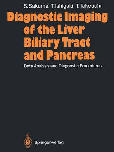
The development and the widespread clinical application of various di agnostic imaging modalities, such as diagnostic ultrasonography, X-ray computed tomography, single photon emission computed tomography, and magnetic resonance imaging, have been beyond all expectation. In particular, ultrasonography and X-ray computed tomography have be come major diagnostic tools for diseases of the liver, the biliary tract, and the pancreas. They often have virtually replaced other conventional imag ing modalities including invasive angiography and percutaneous trans he patic cholangiography. One modality may complement or conflict with another or other modalities. Each modality should be carefully selected with due regard for its diagnostic efficacy. In this book, the first section contains nine chapters dealing with current techniques of each diagnostic modality applicable to the liver, the biliary tract, and the pancreas. The second section deals with diseases of the liver, the biliary tract, and the pancreas and takes the form of case presentation with discussion of the significance of diagnostic imagings and diagnostic procedure. Preparation of the manuscript was made possible by the help of Dr. S. Fujita, who prepared the photographs, and Mrs. Sobajima, who typed the original manuscript. Dr. S. Miura and Miss Y. Shimizu under took the labor of translating our manuscript from Japanese into English. I would like to express my deep appreciation to all these persons, as well as to the contributors to this book, and also to the publishers, Shujunsha, Japan and Springer-Verlag.
$9.00 standard shipping within Australia
FREE standard shipping within Australia for orders over $100.00
Express & International shipping calculated at checkout
This title is printed to order. This book may have been self-published. If so, we cannot guarantee the quality of the content. In the main most books will have gone through the editing process however some may not. We therefore suggest that you be aware of this before ordering this book. If in doubt check either the author or publisher’s details as we are unable to accept any returns unless they are faulty. Please contact us if you have any questions.
The development and the widespread clinical application of various di agnostic imaging modalities, such as diagnostic ultrasonography, X-ray computed tomography, single photon emission computed tomography, and magnetic resonance imaging, have been beyond all expectation. In particular, ultrasonography and X-ray computed tomography have be come major diagnostic tools for diseases of the liver, the biliary tract, and the pancreas. They often have virtually replaced other conventional imag ing modalities including invasive angiography and percutaneous trans he patic cholangiography. One modality may complement or conflict with another or other modalities. Each modality should be carefully selected with due regard for its diagnostic efficacy. In this book, the first section contains nine chapters dealing with current techniques of each diagnostic modality applicable to the liver, the biliary tract, and the pancreas. The second section deals with diseases of the liver, the biliary tract, and the pancreas and takes the form of case presentation with discussion of the significance of diagnostic imagings and diagnostic procedure. Preparation of the manuscript was made possible by the help of Dr. S. Fujita, who prepared the photographs, and Mrs. Sobajima, who typed the original manuscript. Dr. S. Miura and Miss Y. Shimizu under took the labor of translating our manuscript from Japanese into English. I would like to express my deep appreciation to all these persons, as well as to the contributors to this book, and also to the publishers, Shujunsha, Japan and Springer-Verlag.