Readings Newsletter
Become a Readings Member to make your shopping experience even easier.
Sign in or sign up for free!
You’re not far away from qualifying for FREE standard shipping within Australia
You’ve qualified for FREE standard shipping within Australia
The cart is loading…


This title is printed to order. This book may have been self-published. If so, we cannot guarantee the quality of the content. In the main most books will have gone through the editing process however some may not. We therefore suggest that you be aware of this before ordering this book. If in doubt check either the author or publisher’s details as we are unable to accept any returns unless they are faulty. Please contact us if you have any questions.
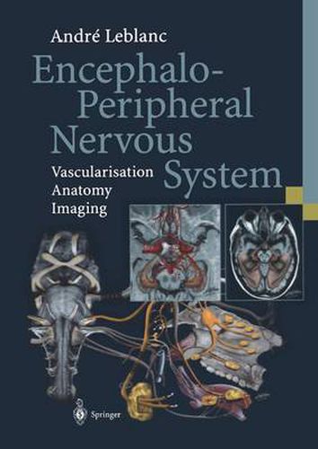
This atlas presents the path taken by each cranial nerve from its real and apparent origins along its intracanalicular and intracranial course to the peripheral organs it supplies. The associated arteries and veins are also described. The author has designed a number of superb original illustrations, including one representing the systematization of the facial nerve with its communicating branches, all the way to the lacrimal and salivary glands, and one depicting the audiovestibular organ in which the walls of the cochlea, vestibule and semicircular canals have been fenestrated so as to visualize the spiral organ of Corti, the utricle, ampullary crests, membranous labyrinth, etc. This excellent practical guide is an invaluable tool for otolaryngologists, anatomists, neurosurgeons, neuroradiologists, radiologists, odontologists, ophthalmologists, neurologists, maxillofacial surgeons, stomatologists and other cranial specialists, as well as for medical students.
$9.00 standard shipping within Australia
FREE standard shipping within Australia for orders over $100.00
Express & International shipping calculated at checkout
This title is printed to order. This book may have been self-published. If so, we cannot guarantee the quality of the content. In the main most books will have gone through the editing process however some may not. We therefore suggest that you be aware of this before ordering this book. If in doubt check either the author or publisher’s details as we are unable to accept any returns unless they are faulty. Please contact us if you have any questions.
This atlas presents the path taken by each cranial nerve from its real and apparent origins along its intracanalicular and intracranial course to the peripheral organs it supplies. The associated arteries and veins are also described. The author has designed a number of superb original illustrations, including one representing the systematization of the facial nerve with its communicating branches, all the way to the lacrimal and salivary glands, and one depicting the audiovestibular organ in which the walls of the cochlea, vestibule and semicircular canals have been fenestrated so as to visualize the spiral organ of Corti, the utricle, ampullary crests, membranous labyrinth, etc. This excellent practical guide is an invaluable tool for otolaryngologists, anatomists, neurosurgeons, neuroradiologists, radiologists, odontologists, ophthalmologists, neurologists, maxillofacial surgeons, stomatologists and other cranial specialists, as well as for medical students.