Readings Newsletter
Become a Readings Member to make your shopping experience even easier.
Sign in or sign up for free!
You’re not far away from qualifying for FREE standard shipping within Australia
You’ve qualified for FREE standard shipping within Australia
The cart is loading…


This title is printed to order. This book may have been self-published. If so, we cannot guarantee the quality of the content. In the main most books will have gone through the editing process however some may not. We therefore suggest that you be aware of this before ordering this book. If in doubt check either the author or publisher’s details as we are unable to accept any returns unless they are faulty. Please contact us if you have any questions.
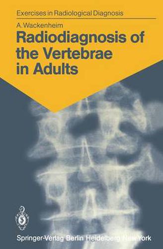
This book is intended for beginners and for those who want to refresh their knowledge of the elementary radioanatomy of the vertebrae, particularly their pathological radioanatomy. I do not pretend, as does Roger Martin du Gard’s hero, that one always must begin with a radiographic examination, but I do believe that a student, especially one interested in radiology, must be able to apprehend an image isolated from its clinical context. To optimize memorization of the image I have selected unmistakable cases with marked, well-evolved lesions. This will enable the studentlater to recognize less distinct images of the same kind. The first section of the book is exclusively iconographic. After studying an image, the reader will find in the second section, under the appropriate reference number, a commen tary illustrated with a realistic drawing by my friend Dr. Csaba Hethalmi. Attention to the following points will assist a fruitful reading: 1. Cases 1-5 involve normal subjects; all other cases are pathological. 2. The reader must imagine that he is conducting a routine examination and draw on his resources to make a practical analysis of an image. As a matter of fact, all the films (except that in case 46, which is the radiograph of a specimen) were -indeed taken under routine conditions using standard pro jections. 3. The cases are in no systematic or nosological order. Each case is illustrated with one, two, or (rarely) three images.
$9.00 standard shipping within Australia
FREE standard shipping within Australia for orders over $100.00
Express & International shipping calculated at checkout
This title is printed to order. This book may have been self-published. If so, we cannot guarantee the quality of the content. In the main most books will have gone through the editing process however some may not. We therefore suggest that you be aware of this before ordering this book. If in doubt check either the author or publisher’s details as we are unable to accept any returns unless they are faulty. Please contact us if you have any questions.
This book is intended for beginners and for those who want to refresh their knowledge of the elementary radioanatomy of the vertebrae, particularly their pathological radioanatomy. I do not pretend, as does Roger Martin du Gard’s hero, that one always must begin with a radiographic examination, but I do believe that a student, especially one interested in radiology, must be able to apprehend an image isolated from its clinical context. To optimize memorization of the image I have selected unmistakable cases with marked, well-evolved lesions. This will enable the studentlater to recognize less distinct images of the same kind. The first section of the book is exclusively iconographic. After studying an image, the reader will find in the second section, under the appropriate reference number, a commen tary illustrated with a realistic drawing by my friend Dr. Csaba Hethalmi. Attention to the following points will assist a fruitful reading: 1. Cases 1-5 involve normal subjects; all other cases are pathological. 2. The reader must imagine that he is conducting a routine examination and draw on his resources to make a practical analysis of an image. As a matter of fact, all the films (except that in case 46, which is the radiograph of a specimen) were -indeed taken under routine conditions using standard pro jections. 3. The cases are in no systematic or nosological order. Each case is illustrated with one, two, or (rarely) three images.