Readings Newsletter
Become a Readings Member to make your shopping experience even easier.
Sign in or sign up for free!
You’re not far away from qualifying for FREE standard shipping within Australia
You’ve qualified for FREE standard shipping within Australia
The cart is loading…


This title is printed to order. This book may have been self-published. If so, we cannot guarantee the quality of the content. In the main most books will have gone through the editing process however some may not. We therefore suggest that you be aware of this before ordering this book. If in doubt check either the author or publisher’s details as we are unable to accept any returns unless they are faulty. Please contact us if you have any questions.
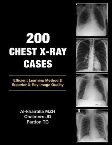
Modern medical practice has seen many advances in imaging over the past ten years. Magnetic Resonance Imaging, CT scanning and Ultrasound investigations have all been added to the repertoire of normal practice. However, the humble chest X-ray remains a crucial first line investigation - particularly for acute medical admissions. Many chest X-rays are requested for a specific purpose e.g. confirmation of pneumonia, but the image may reveal features of previously unsuspected disease of another body system. Digital storage of X-ray images means that a chest X-ray may be viewed at any computer workstation in your hospital. Clinicians now have the opportunity to view these images without waiting for the X-ray packet to be delivered. This can only be advantageous for the patient if the clinician knows what to look for on the image. This book takes you through 200 images in a stimulating manner designed to improve your confidence in reporting the humble chest X-ray.
$9.00 standard shipping within Australia
FREE standard shipping within Australia for orders over $100.00
Express & International shipping calculated at checkout
This title is printed to order. This book may have been self-published. If so, we cannot guarantee the quality of the content. In the main most books will have gone through the editing process however some may not. We therefore suggest that you be aware of this before ordering this book. If in doubt check either the author or publisher’s details as we are unable to accept any returns unless they are faulty. Please contact us if you have any questions.
Modern medical practice has seen many advances in imaging over the past ten years. Magnetic Resonance Imaging, CT scanning and Ultrasound investigations have all been added to the repertoire of normal practice. However, the humble chest X-ray remains a crucial first line investigation - particularly for acute medical admissions. Many chest X-rays are requested for a specific purpose e.g. confirmation of pneumonia, but the image may reveal features of previously unsuspected disease of another body system. Digital storage of X-ray images means that a chest X-ray may be viewed at any computer workstation in your hospital. Clinicians now have the opportunity to view these images without waiting for the X-ray packet to be delivered. This can only be advantageous for the patient if the clinician knows what to look for on the image. This book takes you through 200 images in a stimulating manner designed to improve your confidence in reporting the humble chest X-ray.