Readings Newsletter
Become a Readings Member to make your shopping experience even easier.
Sign in or sign up for free!
You’re not far away from qualifying for FREE standard shipping within Australia
You’ve qualified for FREE standard shipping within Australia
The cart is loading…


This title is printed to order. This book may have been self-published. If so, we cannot guarantee the quality of the content. In the main most books will have gone through the editing process however some may not. We therefore suggest that you be aware of this before ordering this book. If in doubt check either the author or publisher’s details as we are unable to accept any returns unless they are faulty. Please contact us if you have any questions.
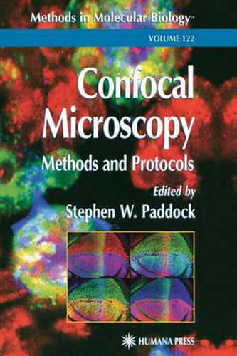
Stephen Paddock and a highly skilled panel of experts lead the researcher using confocal techniques from the bench top, through the imaging process, to the journal page. They concisely describe all the key stages of confocal imaging-from tissue sampling methods, through the staining process, to the manipulation, presentation, and publication of the realized image. Written in a user-friendly, nontechnical style, the methods specifically cover most of the commonly used model organisms: worms, sea urchins, flies, plants, yeast, frogs, and zebrafish. The powerful hands-on methods collected here will help even the novice to produce first-class cover-quality confocal images.
$9.00 standard shipping within Australia
FREE standard shipping within Australia for orders over $100.00
Express & International shipping calculated at checkout
Stock availability can be subject to change without notice. We recommend calling the shop or contacting our online team to check availability of low stock items. Please see our Shopping Online page for more details.
This title is printed to order. This book may have been self-published. If so, we cannot guarantee the quality of the content. In the main most books will have gone through the editing process however some may not. We therefore suggest that you be aware of this before ordering this book. If in doubt check either the author or publisher’s details as we are unable to accept any returns unless they are faulty. Please contact us if you have any questions.
Stephen Paddock and a highly skilled panel of experts lead the researcher using confocal techniques from the bench top, through the imaging process, to the journal page. They concisely describe all the key stages of confocal imaging-from tissue sampling methods, through the staining process, to the manipulation, presentation, and publication of the realized image. Written in a user-friendly, nontechnical style, the methods specifically cover most of the commonly used model organisms: worms, sea urchins, flies, plants, yeast, frogs, and zebrafish. The powerful hands-on methods collected here will help even the novice to produce first-class cover-quality confocal images.